This original research examines whether the interthalamic adhesion, a neural structure at the centre of the brain, and present in only 70% of human subjects, may reduce CRI expansion and contraction movement of the third cranial ventricle
Brain anatomy
It is known that the Third Ventricle (3V) is penetrated bilaterally by the thalamus in approximately 70% of individuals and not present in the remaining 30%.
The key neural integration structure of the brain, the thalamus, has its left and right lobes connected and continuous, passing through the middle of the third ventricle and known as the interthalamic adhesion in 70% of individuals.
By implication, one third of your client list will not possess this structure. Does its presence or absence make a difference to the CRI and can this be palpated?
The commonly seen anatomic map of the deep brain in the sagittal plane, dividing the brain into left and right sides along the mid-line, shows the oval-shaped interthalamic adhesion (IA) in the middle of the third ventricle.
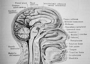 Sagittal plane view of the brain
Few brain sagittal plane illustrations show a 3V without an IA. Your own anatomy text is likely to include an illustration with this standard sagittal plane view, showing an oval IA in the middle of the third ventricle, in the middle of the brain.
 Sagittal plane
When present, this interthalamic adhesion (also known as the massa intermedia) penetrates and forms a continuous neural tissue structure through the middle of the third ventricle lateral walls, connecting the left and right sides of this small hollow organ which has mechanical, neural and hormonal functions.
 Coronal plane of the brain
The existence of an interthalamic adhesion (in 70% of brains) might alter the flexibility of the third ventricle, as the tissue of the IA would be less pliant, and might restrict movement, compared to an open fluid-filled space capable of more expansion, present in only 30% of human brains without an interthalamic adhesion.
Effect on the Cranial Rhythmic Impulse
The subtle pulsatile motion of the Cranial Rhythm is body-wide and includes slow rhythmic changes to the shape and volume of the third cranial ventricle in the midline, at the centre of the head, swelling with expansion and deflating with contraction.
CRI motion occurring in this evolutionarily ancient area of the brain, where mechanical, fluid, neural and hormonal functions all overlap, has a pivotal homeostatic role.
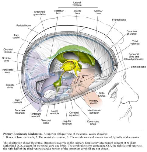 Bone, Fascia and Fluid Systems – Sutherland’s Primary Respiratory Mechanism
The Pituitary and Pineal glands, at the front and back of the 3V respectively (as well as the other Circumventricular Organs on the top of the 3V, in between the pituitary and pineal) are all in motion as the cranial rhythm fluctuates like a tide.
A client who has a third ventricle without an interthalamic adhesion could be capable of greater cranial rhythm amplitude (the range between expansion and contraction) than a client with it present.
What exact effect this anatomical variation has on the CRI, and on still point induction success rates for those individuals with or without an interthalamic adhesion, is a matter of speculation.