When visiting the History of Medicine Museum in Varna, Bulgaria in 1995, I saw an old skull that was quite remarkable. This skull is proof that at least one person from ancient times knew an important anatomical fact that we overlook today, even with the advances of modern medical science.
In antiquity Varna was called Odessos, not to be confused with the modern-day Ukrainian city of Odessa, further north up the Black Sea coast.
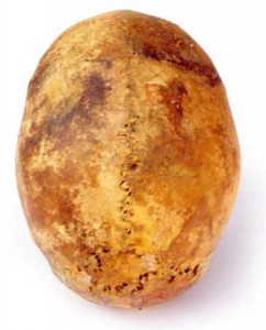
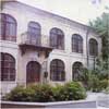
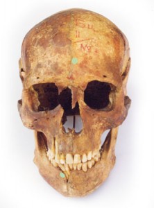
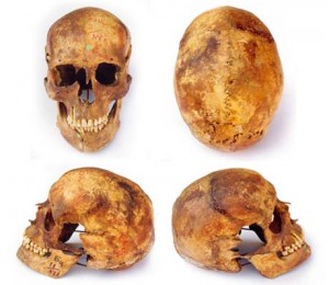
The museum is housed in a picturesque two storey stone building set close to the shore. In a display case with other specimens in the Prehistoric to Bronze Age section on the ground floor was an adult skull with a distinct and telling difference – the sphenoid bone of this skull had been removed, leaving the rest of the cranial bones undisturbed. The museum curator assured me that the skull had not been tampered with since it had been unearthed (date unknown to me) and was presented for display as it had been found. She thought it to be of local origin and to be reliably dated at between 3000 and 8000 years old. Why is this so important?
Whoever removed that sphenoid bone in the Odessos region thousands of years ago knew that it was possible to do so, that is, that the cranial bones DO NOT in fact fuse together, as modern anatomy texts state.
The significance of the Odessos skull is that it extends further back in time the date when evidence exists for ancient knowledge of moveable skull bones.
Pick up any anatomy textbook and you will find it stated that the bones of the head keep fusing together in early life, forming a rigid structure by 18 years of age. This tenet of Western anatomy was not shared by Renaissance Italian anatomy or modern cranial osteopathy How it is possible that such a fundamental discrepancy in our anatomy knowledge has occurred and been perpetuated?
The mobility phenomenon called the Cranial Rhythm can be palpated throughout life, from birth to death. Prior to the time of supposed fusion, it is acknowledged that the cranial bones are indeed separate. The four large gaps between them at birth and in infancy are called fontanelles and are easily observed and felt. The reality is that while the sutures between the individual bones do continue to ossify throughout life, slight mobility between them should remain present, unless trauma or secondary compensation have create restrictions. Individuals in their ninth decade still have a slight mobility between cranial bones that is palpable to the trained therapist.
This is not a dry scientific, anatomic or historical issue only however, as cranial mobility and the associated clinical implications are of vital importance to our health care needs now. To give but one example, it is tragic that motor accident survivors who have suffered cranial trauma (to the extent that a displacement of cranial tissues is visually obvious) are denied appropriate treatment because it is routinely taught that there is no possibility of such bony displacement to occur.
This is especially intriguing since the work of William Garner Sutherland, the American osteopath who hypothesised cranial mobility in 1899 and taught this approach from the 1940’s onwards. While there is now a wealth of anatomic, physiologic, diagnostic and clinical evidence since his time to support this notion, the idea has not permeated orthodox science education.
Imagine viewing an intact skull from the front, looking directly at the nose and eye region. What do you see? As well as the adjacent bones, you see the sphenoid bone. The inside of the cranium is not visible in a normal intact skull because the natural placement of the central sphenoid bone blocks such a view. What I saw in Varna was the inside of that skull clearly visible from the front.
When I looked through the nasal fossa I could see clear through to the internal surface of the occiput bone at the back of the cranium, so it seemed like the skull was completely hollow. The greater wings of the sphenoid on each side were removed, so a space on each side of the skull was present in the area that we commonly call the temples. This layperson term is confusing, as nearby in the skull there are two bones, one on each side of the head, each called a Temporal bone. As the orbital surfaces of the sphenoid (that make up the eye orbits) were missing, there appeared to be larger spaces than usual in the eye region.

This hollow skull in the History of Medicine Museum prompts several questions, as it does not sit comfortably with the orthodox history of anatomy and osteology. Was this removal of the sphenoid a lone local occurrence or was it widely known and practised? Taught even? Exactly how was the sphenoid bone removed without damaging the surrounding bones thousands of years ago?
It should be noted that while the 23 articulations of the sphenoid bone ensure difficulty in this regard, the articular surfaces of the bones that I saw surrounding the now-absent sphenoid looked normal. Did this cranial anatomical expertise extend to other fields?
Is it possible that the movement of the individual cranial bones were known of? The presence of the fontanelles in infancy is an observable anatomic clue that may have been understood by people thousands of years ago. These open question regarding ancient knowledge of anatomy and physiology involving the skull (such as Sutherland’s cranial concept) are only underlined by the implications of this evidence.
The skull with the sphenoid removed that I observed was red in colour – pigmented after death for mystical significance. As to what connection there may be between the red colouring regarded as magical and used in burial ritual, and the removal of the sphenoid, I can only speculate at this time. The Historical Manuscripts section in the museum library may provide some answers, but as the old books would be in the Cyrilliac language, collaborative local research help is needed.
Malcolm Hiort
October 1997 (revised 2013)
FURTHER READING:
Magoun, H I Osteopathy in the Cranial Field, Journal Printing Co., Missouri, 1976.
Retzlaff, E W The Cranium and its Sutures, Springer Verlag, Berlin, 1987.
Sutherland, W G Teachings in the Science of Osteopathy, Rudra, 1990.
Upledger, J E and Vredevoogd, J Craniosacral Therapy, Eastland, Seattle, 1983.


